How Many Lumbar Vertebrae Are In The Vertebral Column – The collection of bones stacked on top of each other to form the spine is known as the vertebral column. Each of these bones is called a vertebra (plural: vertebrae).
The vertebral column has five distinct regions where the vertebrae share distinct characteristics that allow them to form specific joints and generate movements. Each of these fields is assigned a letter and a number, as described below.
How Many Lumbar Vertebrae Are In The Vertebral Column
:max_bytes(150000):strip_icc()/GettyImages-87293476-56e435703df78c5ba0571226.jpg?strip=all "How Many Lumbar Vertebrae Are In The Vertebral Column")
You may also be interested in our Anatomy flashcards collection which contains over 2000 anatomy flashcards in addition to advanced features like spaced repetition.
Anatomy Of The Spine
Before we describe each of these regions and some examples of unusual vertebrae, we will consider the similarities in their basic structure.
Nearly all vertebrae in the human vertebral column have a vertebral body. The vertebral body becomes larger and provides more stability as the vertebral column moves down from the cervical region. This is, in part, due to the increased mass on each vertebral body.
The vertebral body is surrounded by cortical (compact) bone. Within the cortical bone is a dense network of trabecular bone. This trabecular bone is important for hematopoiesis, or blood cell production, as it contains red bone marrow.
The vertebral bodies of two adjacent vertebrae are connected by intervertebral discs. These discs help reduce shock as forces are transmitted through the vertebral column and contribute to improving the range of motion between the vertebrae.
Biomechanics Of The Spine: The Rom Of The Spine
They help to form the lateral margin of the spinal canal, and give rise to four important structures:
The vertebral arch forms the posterior edge of the spinal canal. When the lamina and pedicles are referred to collectively, they are known as the vertebral arch.
Articular processes are small projections of bone extending from the junction between the pedicles and laminae. This process may extend superiorly to form the superior articular process, or extend inferiorly to form the inferior articular process.

These articular processes have articular facets at the tips, which form a (zygapophyseal) articulation with adjacent vertebrae to improve stability within the vertebral column.
Normal Lateral Spinal Column Anatomy
The transverse processes of the vertebral column are bony projections that arise from the same location as the articular processes and travel laterally.
They serve different purposes in different areas of the spine, such as carrying blood vessels or creating attachment sites for ribs.
The spinous processes are postero-inferior projections of bone that exit the vertebral arch posteriorly from the midline. They help limit movement in certain areas of the spine and are the attachment points of many ligaments.
Before introducing each vertebral region, we will explore the different joints in the vertebral column. Here, we focus on common joints in each area.
Amazon.com: Gpi Anatomicals
The lower surface of the vertebral body is attached to an intervertebral disc, which is then attached to the upper surface of the vertebral body beneath it.
On each vertebra, there are two superior articular processes with facets and two inferior articular processes with facets. These form the zygapophyseal, or facet, joints of the vertebral column.
For example, the facets in the thoracic spine have an anterior-posterior orientation, which allows lateral movement but limits flexion and extension.

The anterior and posterior longitudinal ligaments form a long line, which runs from the cervical region to the sacrum. They attach to the anterior and posterior surfaces of the vertebral bodies, respectively. Their function is to limit excessive extension and flexion of the vertebral column respectively
Spinal Anatomy And Back Pain
The ligamentum flavum attaches between the lamina of each vertebra. This is a very thick and strong ligament that limits excessive flexibility of the spine.
The supraspinous ligament runs from the spinous process to the spinous process in a line down the spinal column. This limits excessive flexibility of the spine.
Now that we have covered the general characteristics of most typical vertebrae, we will discuss each vertebral region and highlight some of the characteristics that make the bones of that region particularly recognizable. Check out these three images of the cervical, thoracic, and lumbar spine to see the differences we’ll discuss as we proceed.
Figure 5. Cervical vertebrae for comparison to the thoracic and lumbar vertebrae. Note that this cervical vertebra does not have the bifurcated spinous process of a normal cervical vertebra.
Spinal Cord & Column
The human vertebral column consists of seven cervical vertebrae. They are the most superior vertebral column group, and have several recognizable characteristics which are explained in more detail in the atypical vertebrates article:
There is a separate transverse foramen (hole) in the transverse foramen in the cervical region. These transverse foramina allow the vertebral arteries to pass up the side of the vertebral column before entering the skull through the foramen magnum.
The vertebral arteries usually enter the transverse foramen of C6-C1, but may occasionally pass through the C7 transverse foramen.

Figure 5. Note that this cervical vertebra does not have the bifurcated spinous process of a normal cervical vertebra.
How The Spine Is Organized And Why It Can Hurt: Basic Lumbar Spine Mechanics (8 Minute Read) — Structural Elements
The human vertebral column consists of twelve thoracic vertebrae. They serve as dorsal attachment sites for the ribs, form most of the lateral margins of the thoracic cavity (thorax) and line the thoracic inlet and outlet.
Each typical thoracic vertebra has two hemi-facets (half facets) located on the upper and lower lateral aspects of the thoracic vertebral body. These are positioned so that each of the adjacent thoracic vertebrae is joined by a common rib.
Each distinct rib will be joined by a single transverse process to improve the stability of the ribs and thoracic cage.
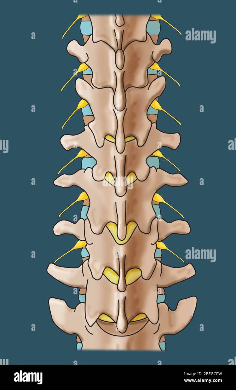
The lumbar vertebra is the last of the individual vertebrae in the vertebral column. There are five lumbar vertebrae, and each provides significant support to the mass of the body above vertebral level. As a result, they have the largest vertebral bodies.
Vertebrae: The Bones Of The Spinal Column
Only a handful of spinal nerves remain to branch from the spinal cord in the lumbar region, and the lumbar vertebrae have a small triangular vertebral foramen.
The sacrum is formed by 5 fused vertebrae that form an inverted triangle, with the base located above the apex.
Co1 and Co2 are always present, but Co3 may be absent or separated from the other two cerebellar vertebrae.

There are no vertebral arches in the coccygeal vertebrae, and thus, no vertebral canal can form in this area.
Understanding The Levels Of The Spinal Cord
Figure 8. Illustration of the sacrum and coccyx as they sit at the base of the vertebral column along with the sacroiliac joint.
Scoliosis refers to a lateral curvature of the spine that can occur in any vertebral region, but most commonly occurs in the thoracolumbar spine.
It may be caused by improper posture due to muscular or skeletal abnormalities and can occur at any age. It affects women more than men.
Signs and symptoms depend on the location, but pain is common. In thoracic scoliosis, dyspnea and restrictive lung function patterns may be seen. In lumbar scoliosis, abdominal pain and ‘constriction’ of the abdominal viscera are common.
Vertebral Column Anatomy: Cervical, Thoracic, Lumbar, Sacral Spine — Ezmed
Conservative therapy with physiotherapy, braces and exercise management may be curative in mild disease. More advanced disease may require surgical intervention.
Figure 9. A series of photographs showing scoliosis. Far left: Scoliosis before treatment. Note the lateral curvature of the thoracolumbar spine and discrepancy in interscapular height. Middle left: A brace used to treat scoliosis. Middle right: X-ray showing lateral curvature of the spinal column. Far right: X-ray after treatment.
Kyphosis usually refers to pathological and excessive flexion of the primary spinal curvature: thoracic or sacral curvature.

Thoracic kyphosis manifests with heavy slouched posture and back pain. It is usually a product of low bone mineral density that causes osteopenic or osteoporotic crush fractures of the thoracic vertebrae.
Lumbar Spine Injury
Lordosis refers to the pathological and excessive extension of the secondary spinal curves: cervical or lumbar curvature.
Lumbar lordosis presents with anteriorly rotated hips and excess extension of the lumbar spine (“hollow back”, or “saddleback”).
Common causes of lumbar lordosis can be tight hip flexors and erector spinae muscle groups, pregnancy, and puberty growth spurts.
Figure 10. Note the excessive anterior curvature of the vertebral column in the patient on the right, causing his posture to be stooped.
Lumbar Vertebra L3
The upper half of the intervertebral disc sends arranged sheets of type I collagen fibers to attach to the base of the superior vertebral body; And the lower half of the intervertebral disc sends these organized sheets to the top of the lower vertebral body.
Thus, one point of weakness in the intervertebral disc is this (perpendicular) middle portion of the disc, where the disorganized type I collagen fibers occur. The weakest part of the intervertebral disc is in the middle of the posterolateral portion, where the disc material can slip and spill into the vertebral canal causing spinal cord or nerve compression.
The most common locations for disc herniation are the lower cervical region and lower lumbar region. In both areas, the primary complaint is shooting or spreading pain when the spine or limb is moved. Later in the course of the disease, motor dysfunction may occur.

To assess for disc herniation in the lumbar region, the supine leg raise test is completed. Regeneration of symptoms in the first 30 degrees of hip flexion is a positive sign, and suggests the need for further workup for lumbar disc herniation.
Amazon.com: Jujne Human Spine Model, Life Size Lumbar Vertebrae Spine Set Anatomy Model Consists Of 5 Lumbar Vertebrae With Intervertebral Discs Lumbar Nerves And Spinal Cord
Figure 11. Four stages of disc herniation. Depending on the severity of the injury, any
Vertebrae in lumbar spine, vertebral column in humans, how many vertebrae make up the vertebral column, vertebral column vertebrae, pain in the vertebral column, pain in lumbar vertebrae, the lumbar vertebrae, vertebrae in the vertebral column, vertebrae in vertebral column, lumbar vertebral column, bones in the vertebral column, how many vertebrae in vertebral column

Most Common Cause Of Low Platelet Count

How To Get Rid Of Bees In Vinyl Siding


