What Is The Function Of The Auditory Ossicles – Abdominal wall Peritoneum Stomach Spleen Liver Pancreas Small intestine Large intestine Kidneys and bladder Abdominal nerves, vessels, and lymph nodes
Pelvic Girdle and Floor Female Pelvis and Reproductive Organs Male Pelvis and Reproductive Organs Bladder and Urethral Tract Perineum Pelvic Nerves, Vessels, and Lymph Nodes
What Is The Function Of The Auditory Ossicles

Overview Skull Face and skull Infratemporal region and pterygopalatine fossa Orbit and contents Nasal region Ear Oral cavity Teeth Throat Neck
How Does Hearing Work?
Overview Cerebrum Diencephalon Cerebrum Brainstem Meninges, ventricular system, and subarachnoid space Blood supply to the brain Spinal cord Nervous system pathways Cranial nerves Peripheral nervous system
Cardiovascular System Nervous System Associated System Musculoskeletal System Respiratory System Urinary System Endocrine System Digestive System Lymphatic System Male Reproductive System Female Reproductive System
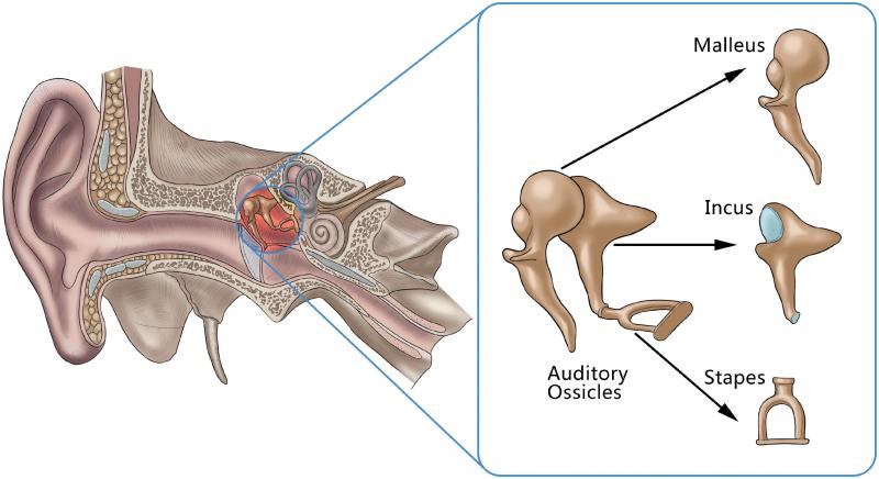
In this article we will discuss the auditory bones, namely malleus, incus and stapes. Inside the middle ear are the smallest bones in the body – the auditory ossicles or ear bones. By definition, these three bones are named after their shape: malleus (“hammer”), incus (anvil), and stapes (stirrup). During development, the auditory ossicles are the first bones to fully ossify and mature at birth, where they do not grow.
Bones are located in the middle ear and are suspended by ligaments. They articulate with each other through synovial joints to form a chain along the middle ear from the tympanic membrane (laterally) to the oval window (medially). Bones transmit mechanical vibrations of the tympanic membrane through this chain to the oval window, where fluid in the inner ear moves and excites receptors. This process allows sound to be converted into electrical signals that are sent to the brain. This article will explore the function of the auditory ossicles, their skeletal features, joints, associated muscles and some clinical aspects.
Anatomy And Physiology: External Ear
The most lateral and largest of the auditory bones is the malleus (hammer). It has several parts: handle, spartate process, lateral process, frontal process, neck and head.
The handle of the malleus (also called the manubrium) is a downward extension embedded in the medial surface of the tympanic membrane. As the handle extends downward, it narrows. At the end of the handle is the spatulate process of the malleus, which connects to the pars tensa of the tympanic membrane with ligaments. This attachment pulls the tympanic membrane medially and forms a depression in the tympanic membrane known as the umbo. Superior to the handle, the lateral process projects laterally as a slight cone at the root of the handle. It is attached to the superior part of the tympanic membrane by the anterior and posterior malleolar folds. The anterior process is much longer than the lateral process. Superior to the lateral process and inferior to the neck, it projects anteriorly like a coil and attaches to the anterior wall of the middle ear. The anterior process is also known as the Folian or Rau process.
Superior to the lateral and frontal processes is the neck of the malleus. It is quite narrow and lies on top of the pars flaccida of the tympanic membrane. Superior to the neck is the rounded head of the malleus, which sits in the epitympanic depression. On its posterior side, it articulates posterolaterally with the body of the incus by a small synovial joint: the incudomalleolar joint.

The incus is the middle auditory bone. It is suspended medially to the malleus and laterally to the stapes and connects these bones with synovial joints. It consists of: body, short limb, long limb/process and lens process.
Ligaments Of Auditory Ossicles
The body of the incus articulates endoscopically with the head of the malleus. Like the head of the malleus, it sits in the epitympanic depression. The short leg projects posteriorly from the body and is the attachment point for the posterior ligament of the incus. The long leg/process projects inferiorly and anteroposteriorly, parallel to the handle of the malleus. At its lower end, it bends at an angle of 90° and forms the lens process, which articulates with the head of the stapes by the incudostapedial joint.
The most medial and smallest auditory bone is the stapes. Recognizable features on stapes are the head (capitulum), forelimb, hindlimb, and foot (footplate).
On its lateral side, the head of the stapes (capitulum) articulates with the lens process of the long limb of the incus at the incudostapedial joint. The front and rear limbs emerge from the head and connect to the oval-shaped base. The base of the stapes (footboard) then sits in the oval window of the labyrinthine (middle) wall of the tympanic cavity.
Two skeletal muscles are attached to the auditory ossicles that contract in response to loud sounds. This process reduces the movements of the auditory ossicles, thereby reducing the vibrations caused by loud sounds to protect the structures of the inner ear. These two muscles are the tensor tympani (eustachian) muscle and the stapedius muscle.
The Septum Configuration In The Chinchilla Middle Ear Cavity. A Lateral…
The tensor tympani muscle lies in a bony canal superior to the pharyngotympanic (Eustachian) tube and passes posteriorly to insert onto the handle of the malleus. When it contracts, it pulls the handle of the malleus medially, which puts tension on the tympanic membrane. Since the tympanic membrane is under tension, it is unable to vibrate as much and the amplitude of oscillation is reduced. This is done in response to loud noises to protect the structures of the inner ear.
Superior aspect of the cartilaginous part of the pharyngotympanic (Eustachian) tube; Large wing of the sphenoid bone; Bony canal of the tensor tympani muscle of the petrous part of the temporal bone
The stapedius muscle attaches to the inside of the pyramidal eminence on the mastoid (posterior) wall of the middle ear, passes through a foramen at the top of the pyramidal eminence, passes anteriorly and enters the back of the neck. stapes. As it contracts, the stapes is pulled back and its base leans into the oval window. This action tightens the annular ligament of the stapes, reducing oscillation and preventing excessive movement of the stapes. Like the tensor tympani muscle, the stapedius muscle contracts in response to loud sounds.

The auditory ossicles transmit the vibrations of the tympanic membrane to the oval window through the middle ear. At the oval window, a wave is generated to move the fluid in the inner ear, which excites receptor cells and allows these mechanical vibrations to be converted into electrical signals. Because the base of the stapes (attached to the oval window) is much smaller than the tympanic membrane, the shock forces at the base are 10 times greater than those of the tympanic membrane. This means that the bones increase the vibration force but the amplitude decreases as the vibrations are transmitted through each bone. This converts large-amplitude, low-power shocks into small-amplitude, high-power shocks.
Auditory Transduction And Pathways: Video & Anatomy
Passive learning methods like reading and re-reading are very ineffective! Learn the anatomy of the ear faster and more efficiently using s labeling ear diagrams and quizzes!
When we hear something, this sound must be converted from sound waves into electrical signals for the brain to process. First, sound waves enter the external acoustic meatus or auditory canal and vibrate the tympanic membrane located at the end of this canal. On the opposite side of the tympanic membrane, on the medial side, the malleus is attached with its handle. This is where a series of movements of the auditory ossicles begin.
First, movements of the medial part of the tympanic membrane also move the handle of the malleus medially. This, in turn, moves the head of the malleus laterally, which moves the head of the incus laterally, because the head of the malleus and the head of the incus articulate with each other. As the head of the incus moves laterally, its long process moves medially. As the long process of the incus articulates with the stapes, the stapes also moves medially. The base of the stapes is attached to the oval window, so medial movement of the stapes means that the oval window also moves medially. Movement of the oval window generates a wave in the fluid-filled inner ear that stimulates receptor cells to transmit electrical signals to the brain via the cochlear portion of the vestibulocochlear nerve (CN VIII).
Osseous chain dysfunction occurs when the auditory bones do not articulate correctly: they either fuse together and lose free movement; Or they are too far away to transmit sound to the oval window. There are a number of causes, including trauma, infection, malformations of the bones from birth, otosclerosis (abnormal growth of the bones) or chronic suppurative otitis media (inflammation). Ossicular chain discontinuity is usually treated with a hearing aid or ossicular chain reconstruction surgery.
Ossicles: Anatomy And Functions
Paralysis of the tensor tympani or stapedius muscle makes it impossible to reduce the amplitude and oscillation created by loud sounds. This causes a hypersensitivity to loud sounds called hyperacusis. Paralysis of these muscles is caused by damage to the nerves that supply them. This type of injury
Function of the external auditory meatus, what is the function of auditory nerve, function of the ossicles, function of auditory tube, function of auditory nerve in ear, function of the auditory ossicles, function of the auditory canal, function of the auditory nerve, what is the function of the ossicles, what is the function of the ear ossicles, function of the auditory nerve in the ear, what is the auditory ossicles

How Many White Blood Cells Are In The Human Body

Symptoms Of Low Testosterone In Older Males


