What Is The Function Of Elastic Connective Tissue – Dense connective tissue contains more numerous, thicker and denser fibers than loose connective tissue, but is much less cellular than cellular. Types of Dense Connective Tissue Dense Regular Connective Tissue Dense Irregular Connective Tissue Elastic Connective Tissue
Parallel bundles of collagen fibers with fibroblasts between collagen fibers White, tough, flexible (forms tendons) Also called white fibrous connective tissue.
What Is The Function Of Elastic Connective Tissue

Dense irregular connective tissue contains irregularly arranged collagen fibers in areas of the body where tension occurs in different directions. formed on sheets such as the dermis of the skin. heart valves, perichondrium – the tissue surrounding the cartilage, periosteum – the tissue surrounding the bone.
Solved This Tissue Is [select] . It Has Collagen And Elastic
Collagen fibers are irregularly arranged (weave) Tissue can withstand tension in any direction Very tough tissue — white of eyeball, dermis
Branched elastic fibers and fibroblasts Lung tissue, vocal cords, and ligaments between vertebrae stretch and return to their original shape.
6 Cartilage cartilage consists of a dense network of collagen fibers and elastic fibers embedded in chondroitin sulfate. The strength comes from collagen fibers and the flexibility comes from chondroitin sulfate. It is surrounded by a dense irregular connective tissue membrane called the perichondrium. Unlike other connective tissues, cartilage is devoid of blood vessels and nerves (except for the perichondrium).
7 Cartilage growth occurs by intermediate growth (expansion from the inside) and compound growth (from the outside). Types of Cartilage Hyaline Cartilage Cartilage Elastic Cartilage
Tissues, Part 2: Connective Tissue
Hyaline cartilage (Table 4.4G) is the most abundant, but weakest, with fine collagen fibers embedded in a gel-like matrix. Table 4.4H) contains bundles of collagen fibers in the matrix, which is the strongest of the three types of cartilage, lacks perichondrium, and is located between the vertebrae (intervertebral discs) and the pubic symphysis.
Perichondrium, which contains a thread-like network of flexible fibers Provides strength and elasticity Maintains the shape of some organs Forms the outer ear and end of the nose
Chondrocytes are located in spaces called lacunae Repair is very slow because there are no blood vessels or nerves Reduces friction in joints such as articular cartilage

Because they are vascularized, they grow slowly and regenerate Interstitial growth Chondrocytes divide and form new matrix Occurs in childhood and adolescence Chondroblasts secrete matrix at the surface and increase width.
Connective Tissue Supports Tissues And Organs
14 Bone (Bone) Tissue Provides protection, mobility, stores minerals, and places blood cells to form Bone (bone) consists of mineral salts, collagen fibers, and cells called osteocytes. Spongy bone spongy, interstices, trabeculae trabeculae = bony bone surrounded by red marrow, no osteons (cell organization) Compact bone (Table 4.4J) the basic structural unit of hard, compact bone is the osteon (haversian system).
Mineralized matrix calcium and phosphate lamellae (rings)—give it its rigidity, and interwoven collagen fibers provide strength Lacunae are small spaces between layers that contain mature bone cells called osteocytes. Canaliculi are small canals containing osteocyte processes that provide pathways for the transport of nutrients and waste. The central (Haversian) canal contains blood vessels and nerves.
The fluid matrix of blood is called plasma-forming elements (Table 4.4K) Lymph is the interstitial fluid that flows through lymphatic vessels. Contains less protein than blood plasma. Move cells and substances (eg, lipids) from one part of the body to another.
Cell types include red blood cells (erythrocytes), white blood cells (leukocytes), and platelets, which are involved in clotting, immune function, and transport of O2 and CO2.
Solved] Identifying The Functions And Locations Of Loose Connective…
To operate this website, we record and share user data with processors. To use this website, you must agree to our Privacy Policy, including our Cookie Policy. As the name suggests, one of the main functions of connective tissue is to connect tissues and organs. In contrast to epithelial tissue, which consists of tightly packed cells with little or no extracellular space between them, connective tissue cells are dispersed throughout the matrix. The matrix usually contains large amounts of extracellular material derived from the connective tissue cells embedded within. The matrix plays a key role in the function of this tissue. The main component of the matrix is usually a powdery substance criss-crossed by protein fibers. This ground substance is usually liquid, but can be mineralized and hard, like in bone. Although connective tissues come in many different forms, they generally share three common characteristics: cells, large amounts of amorphous powder, and protein fibers. The size and structure of each component is related to the function of the tissue, from the solid powder of bone that supports the body to the inclusion of specialized cells; for example, a phagocytic cell that engulfs pathogens and rids the tissue of cellular debris.
Connective tissue has many functions in the body, but most importantly it supports and connects other tissues; from the connective tissue sheaths that surround muscle cells, to the tendons that attach muscles to bones, to the skeleton that supports body posture. Protection is another major function of connective tissue in the form of fibrous capsules and bones that protect delicate organs and, of course, the skeletal system. Special connective tissue cells protect the body from invading microorganisms. The transport of fluids, nutrients, wastes and chemicals is provided by specialized fluid connective tissues such as blood and lymph nodes. Fat cells store excess energy in the form of fat and contribute to thermal insulation of the body.
All connective tissues arise from the mesodermal layer of the embryo. The first connective tissue formed in the embryo is mesenchyme, the stem cell line from which all connective tissues later develop. Clusters of mesenchymal cells are distributed throughout adult tissues and provide cells necessary for repair and regeneration after connective tissue injury. The second type of embryonic connective tissue is formed inside the umbilical cord and is called mucous connective tissue or Wharton’s jelly. This tissue is no longer present after birth, leaving only mesenchymal cells scattered throughout the body.

The three broad classes of connective tissue are classified according to the properties of their parent material and the type of fibers contained in the matrix (Table 1). Connective tissue includes loose connective tissue and dense connective tissue. Both tissues contain a variety of cell types and protein fibers suspended in a viscous powder. Dense connective tissue is supported by bundles of fibers that provide tensile strength, flexibility, and protection. In loose connective tissue, fibers are loosely organized, leaving large spaces between them. Supporting connective tissue and cartilage provide structure and strength to the body and protect soft tissue. Several distinct cell types and tightly packed fibers in the matrix characterize these tissues. In bone, the matrix is hard and is defined as calcified due to the accumulated calcium salts. In connective tissue, that is, lymph and blood, various specialized cells circulate in an aqueous fluid containing salts, nutrients, and dissolved proteins.
Connective Tissues Diagram
Fibroblasts are found in all connective tissues (Figure 1). Fibrocytes, adipocytes, and mesenchymal cells are persistent cells, meaning they remain in the connective tissue. Other cells move in and out of the connective tissue in response to chemical signals. Macrophages, mast cells, lymphocytes, plasma cells, and phagocytic cells are found in connective tissue, but are actually part of the immune system that protects the body.
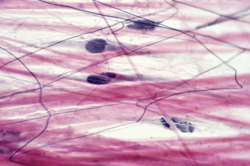
Figure 1. Connective tissue correct. Fibroblasts form this fibrous tissue. Connective tissue includes fibrocytes, adipocytes, and mesenchymal cells. LM × 400. (Photomicrograph courtesy of the Regents of the University of Michigan Medical School © 2012)
The most abundant cells in connective tissue are fibroblasts. Polysaccharides and proteins secreted by fibroblasts combine with the extracellular fluid to form a sticky powder, which, together with absorbed fibrous proteins, forms the extracellular matrix. As you might expect, fibrocytes, the inactive form of fibroblasts, are the second most common cell type in connective tissue.
Adipocytes are cells that store lipids as droplets that fill most of the cytoplasm. There are two main types of fat cells: white and brown. Brown fat cells store a few drops of lipids and are highly metabolically active. In contrast, white adipose fat cells store lipids as one large droplet and are less metabolically active. Their effectiveness in storing large amounts of fat is also witnessed in obese people. The number and type of adipocytes are tissue and location specific and vary among individuals in the population.
Part 3 Tissues.
Mesenchymal cells are the multipotent stem cells of adults. These cells can differentiate into all types of connective tissue cells needed to repair and heal damaged tissue.
Macrophages are large cells derived from monocytes, a type of blood that enter the connective tissue matrix of blood vessels. Macrophage cells are an important component of the immune system, which protects the body against potential pathogens and degraded host cells. When stimulated, macrophages release cytokines and produce small proteins that act as chemical messengers. Cytokines recruit other cells of the immune system to the site of infection and stimulate their activity. Roaming or free macrophages move rapidly with amoeboid motility and engulf infectious agents and cellular debris. In contrast, resident macrophages are permanent residents of tissues.
Mast
:max_bytes(150000):strip_icc()/dense_connective_tissue-56a09aee3df78cafdaa32ca1.jpg?strip=all "What Is The Function Of Elastic Connective Tissue")
What is the function of dense connective tissue, what is the function of areolar connective tissue, what is the main function of connective tissue, what is the general function of connective tissue, what is the function of elastic tissue, what is the primary function of connective tissue, elastic connective tissue function and location, what is elastic connective tissue, function of the connective tissue, what is the function of connective tissue, what is the function of adipose connective tissue, elastic connective tissue function

The Effects Of Climate Change On Agriculture

Definition Causes And Effects Of Global Warming


