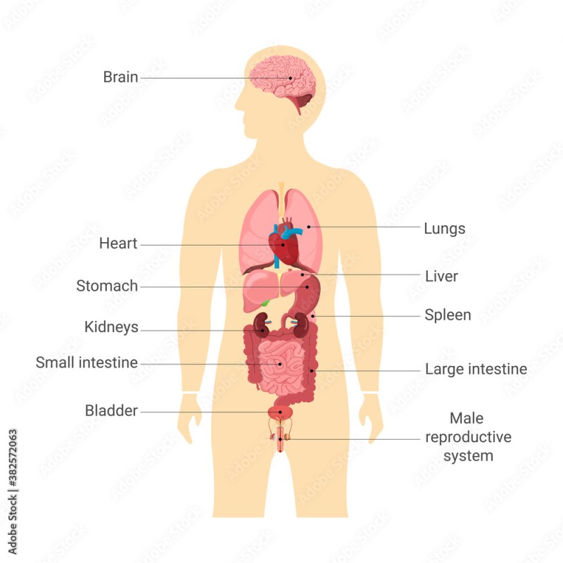
Labeled Diagram Of Internal Organs Human Body – This chart provides a simple, easy-to-understand overview of the location and functions of the body’s major internal organs, including the heart, lungs, stomach, kidneys, diaphragm, spleen, liver, pancreas, large and small intestines, gallbladder, and brain. Ideal for patients and students.
Anatomical diagram of the human skull shows the front and lateral aspects of the skull, the base of the skull (including the inner surface), the sagittal section through the skull, the horizontal section through the maxilla,…
Labeled Diagram Of Internal Organs Human Body

This detailed diagram shows a clear anatomical view of the hair within the skin and the hair shaft. It also explains scalp hair types and hair fiber characteristics. Hair loss appears…
Appendix Diagram Images, Stock Photos, 3d Objects, & Vectors
Anatomical diagram of the organs of hearing and balance shows the anatomy of the ear including the right ear, right tympanic membrane, middle ear, auditory ossicles, membranous labyrinth, diaphragm…
The human body diagram is a comprehensive, highly detailed and brilliantly illustrated presentation of the major sectors and functions of a living human. Among them is the muscular system and its most important elements.
This informative diagram shows a variety of views of a normal prostate, including sagittal, posterior, superior, and anterior. It also shows areas of the prostate, blood vessels and innervation. Hormonal…
Learn all about the endocrine system with this beautifully illustrated wall chart from Body Scientific International. It includes the pituitary gland, thyroid gland, pituitary gland, and pituitary gland.
Human Anatomy Female Body And Organs Diagram Stock Illustration
Learn all about the digestive system with Body Scientific International provides you with a diagram of the digestive system depicting close-ups of the oral cavity, stomach, liver, gallbladder, pancreas…
The human vascular system anatomy diagram is a complete representation of the vein system that runs throughout our bodies. The chart is beautifully drawn and labeled in great detail. It includes…
Dermatomeschart provides an easy-to-understand map of the approximate areas of skin supplied by nerves from a single spinal root. The central figure is beautifully and colorfully illustrated…

Stunning illustrations from Body Scientific International depict the general anatomy of the urinary system in this wall chart. There are also illustrations of the kidneys and nephron filtration…
Internal Structure Of The Human Body. Internal Organs Designations.
The Human Muscular System Anatomy Chart is a wonderful, complete guide to the human muscular system, showing a human figure from the front and back. Each aspect is carefully labeled, and…
The Human Skeletal Anatomy Diagram shows three views of the human skeleton (anterior, posterior, and lateral) and has been very carefully labeled and drawn, resulting in one of the most beautiful and impressive drawings… Download this vector illustration of the digestion process called Internal Organs Vector Educational Diagram now . And search more of iStock’s library of royalty-free vector art graphics featuring human digestive system graphics available for quick and easy download.Product #:gm1243413609 $12.00 iStock In Stock
Digestion vector illustration internal organs educational infographic labeled. Digestive system physiology and anatomical explanation. Elements of the digestive system on a simple silhouette of the human body.
Royalty-free licenses let you pay once to use copyrighted images and video clips in personal and commercial projects on an ongoing basis without requiring additional payments each time you use that content. It’s a win-win, and it’s why everything on iStock is only available royalty-free — including all human digestive system images and footage.
Internal Organs Chart
Royalty-free licenses are the best option for anyone who needs to use stock images commercially, which is why every file on iStock — whether it’s a photo, illustration or video clip — is only available royalty-free.
From social media ads to billboards, PowerPoint presentations to feature films, you’re free to modify, resize and customize every asset on iStock — including all Human Digestive System images and footage — to fit your projects. With the exception of “Editorial use only” photos (which can only be used in editorial projects and can’t be modified), the possibilities are limitless.
© 2023 LP. iStock design is a trademark of LP. Browse millions of high-quality stock photos, illustrations, and videos. In a multicellular organism, an organ is a group of tissues linked into a structural unit to serve a common function.

In the hierarchy of life, an organ is located between tissues and the organ system. Tissues are made up of cells of the same type that work together in a function. Tissues of various types come together to form an organ with a specific function. For example, the intestinal wall consists of epithelial tissue and smooth muscle tissue.
Solved] Diagram 1: Label Refer To Figure 6 3 In The Textbook And Type Your…
Two or more organs work together to carry out a specific function in the body and form an organ system, also called a biological system or body system.
Organ tissues can be broadly classified as parietal tissue, functional tissue, stroma, and structural tissue with supportive, connective, or auxiliary functions. For example, the gland tissue that makes hormones is the parenchyma, while the stroma includes the nerves that innervate the parenchyma, the blood vessels that oxygenate and nourish it and carry its metabolic wastes, and the connective tissue that provides a suitable place for its growth. It must be present and installed. The major tissues that make up an organ have common embryonic origins, such that they arise from the same germ layer. Organs are found in most multicellular organisms. In single-celled organisms such as eukaryotes, the functional counterpart of the organ is known as an organelle. In plants, there are three main organs.
The number of organs in any organism depends on the definition used. By one widely adopted definition, 79 organs in the human body have been identified.
With the exception of placozoans, multicellular animals including humans have a variety of organ systems. These specific systems are widely studied in human anatomy. The functions of these organ systems often share significant overlap. For example, both the nervous system and the endocrine system operate through a common organ, the hypothalamus. For this reason, the two systems are combined and studied as a neuroendocrine system. The same applies to the musculoskeletal system because of the relationship between the muscular system and the skeletal system.
Human Organs Labelled Stock Illustrations
The abdominal organs can be classified as solid organs or hollow organs. The solid organs are the liver, pancreas, spleen, kidneys, and adrenal glands. The hollow organs in the abdomen are the stomach, intestines, gallbladder, bladder, and rectum.
The term “visceral” contrasts with the term “parietal” which means “of or pertaining to the wall of a body part, organ, or cavity.”
The two terms are often used to describe a membrane or piece of connective tissue, referring to opposite sides.

The relationship between major animal lineages, indicating how long ago these animals shared a common ancestor. On the left, important organs are shown, allowing us to determine how long ago these organs evolved.
Inner Organs Human Anatomy Chart Names Stock Vector By ©furian 258471888
The level of organismal organization in animals can be first discovered in the flatworms and the most derived phyla, the bilateria. Less advanced taxa (such as Placozoa, Porifera, Ctophora, and Cnidaria) do not show fusion of their tissues into organs.
More complex animals consist of different organs that have evolved over time. For example, the liver and heart evolved in chordates about 550-500 million years ago, while the intestines and brain are more recent, having originated in the ancestors of vertebrates, insects, molluscs, and worms about 700-650 million years ago.
Given the ancient origin of most vertebrate organs, researchers have looked for model systems in which organs evolved more linearly and ideally evolved many times independently. One outstanding model for this line of research is the plecta, which has evolved more than 100 times independently in vertebrates, evolved relatively straight in some lineages, and is found in intermediate forms in extant taxa.
Studies of plactite evolution have identified a variety of genetic and physiological processes that contribute to organ origin and development. These processes include the reuse of existing animal tissues, the acquisition of new functional properties, and novel interactions. A distinct type of tissue.
Amazon.com: Monmed Human Torso Model
The flower is the reproductive organ of angiosperms. This hibiscus flower is hermaphrodite, and contains lobes and pistils.
The study of plant organs is covered in Plant Morphology. Plant organs can be divided into vegetative and reproductive. Vegetative plant organs include roots, stems, and leaves. The reproductive organs are variable. In flowering plants, they are present in the flower, seed, and fruit.
In conifers, the organ that holds the reproductive structures is called the cone. In other divisions (phylum) of plants, the reproductive organs are called strobili, in Lycopodiophyta, or simply gametophores in algae. Common nomenclature for the organ system in plants includes differentiation between shoot and root. All aboveground plant parts (in non-epiphytes), including functionally distinct leaf and flower organs, can be classified together as a suborgan system.

Vegetative organs are essential for maintaining plant life. While there can be 11 organ systems in animals, there are much fewer in plants, some of which perform vital functions, such as photosynthesis, while reproductive organs are essential in reproduction. However, if there is asexual vegetative reproduction, the vegetative organs are those that create the new generation of plants (see clonal colony).
Diagram Internal Organs Stock Vector. Illustration Of Healthcare
Many societies have an organ donation system, where an organ from a living or deceased donor is transplanted into a person with a failing organ. Transplantation of larger solid organs often requires immunosuppression to prevent organ rejection or graft-versus-host disease.
Organ transplants began as scientists learned more about anatomy
Diagram internal organs human body, human anatomy internal organs diagram, labeled human body organs, human body internal organs diagram female, labeled diagram of human organs, human internal organs diagram labeled, diagram of the human body showing internal organs, diagram of human internal organs, the human body diagram internal organs, diagram human body organs labeled, human internal organs labeled, internal organs diagram of the human body

What Is The Difference Between Sustainability And Sustainable Development

Mitochondria Is The Powerhouse Of The Cell


